
Correlia
An ImageJ plug-in to co-register and visualise multi-modal correlative micrographs.
Abstract
Correlia is an open-source ImageJ/FIJI plug-in for the registration of 2D multi-modal microscopy data-sets. The software is developed at ProVIS - Centre for Correlative Microscopy and is specifically designed for the needs of chemical microscopy involving various micrographs as well as chemical maps at different resolutions and field-of-views.
Licensing and Download
Correlia is free software
licensed
under the GNU General Public License (GPL), version 3. It is open source and the Java code can be adapted as long as the GPL is respected. Correlia has been tested successfully on MS Windows and Linux systems. Here Correlia is made available via pre-compiled jar archive as well as source code.
Java archive (jar):
a jar archive of Correlia can be downloaded here:
Download correlia.jar (366.2 KB)
Requirements:
- ImageJ 1.52 with bUnwarpj 2.6.12 or
- respective FIJI distribution
The installation is as easy as copying the jar-file to the plugins folder of your ImageJ/FIJI installation on your computer.
Correlia source code:
The source code is available on
git.ufz.de
.
- Florens Rohde, Ulf-Dietrich Braumann, and Matthias Schmidt
"Correlia: an ImageJ plug-in to co-register and visualise multi-modal correlative micrographs"
Journal of Microscopy 280 (1), 3-11 2021
DOI: 10.1111/jmi.12928 - Matthias Schmidt, Florens Rohde, Ulf-Dietrich Braumann
"Visualization and co-registration of correlative microscopy data with the ImageJ plug-in Correlia"
In: T. Müller-Reichert, P. Verkade (eds.)
Correlative light and electron microscopy IV
Methods Cell Biol. 162 (2021)
Elsevier, p. 353-388
DOI: 10.1016/bs.mcb.2020.10.001 - Florens Rohde
"Entwicklung eines nichtlinearen Algorithmus zur automatischen Registrierung korrelativer Mikroskopiedaten"
(Development of a non-linear algorithm for automatic registration of correlative microscopy data-sets)
Master's Thesis (2018)
Faculty of Electrical Engineering and Information Technology, Leipzig University of Applied Sciences (HTWK Leipzig) - Matthias Schmidt, Florens Rohde, and Ulf-Dietrich Braumann
"Correlia: An ImageJ-based Tool for the Co-registration of Multi-modal Correlative Microscopy Data."
In: Microscopy Conference 2019 (MC 2019, Abstracts), page 761, contribution LS5.005.
Deutsche Gesellschaft für Elektronenmikroskopie (DGE), September 2019.
https://epub.uni-regensburg.de/40685/1/MC%202019_Proceedings.pdf
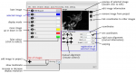
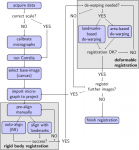
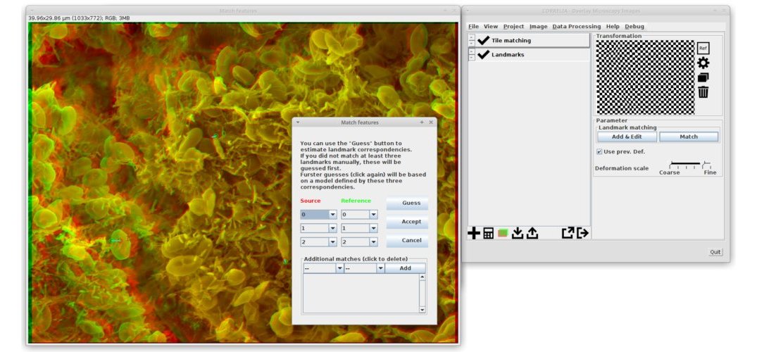
Example 1 - Rigid registration of artificial data
This example should provide an easy start with Correlia. It illustrates Correlia's basic functionality for rigid registration using an artificial data set. The data set contains images are already scaled but are of different pixel sizes and orientation. Please read the pdf file provided with the example.

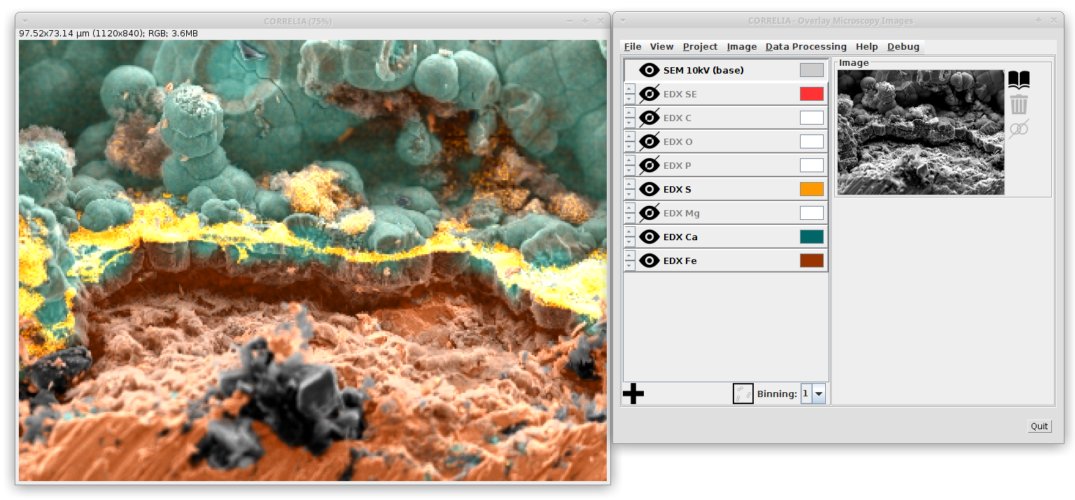
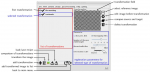
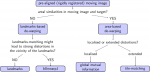
Example 2 - Rigid registration of SEM-EDX data
This example illustrates rigid registration of elemental maps obtained by energy-dispersive X-ray spectroscopy (EDX) onto a scanning electron microscopy (SEM) micrograph. The SEM micrograph are already calibrated but the EDX data is without a scale bar. Before starting Correlia FIJI tools have to be employed in order to put a scale to the EDX maps.

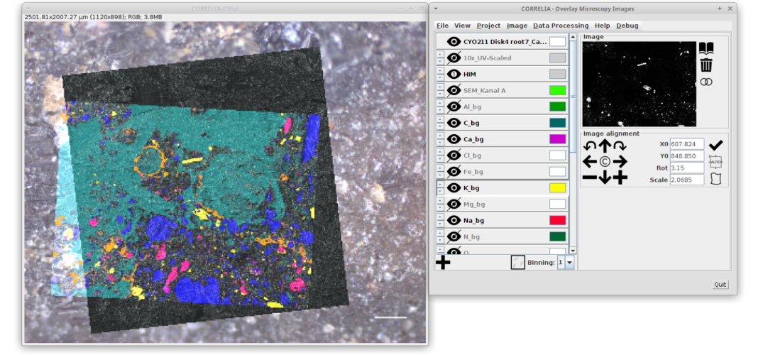
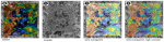
Example 3 - Effects of Surface Roughness and Mineralogy on the Sorption of Cm(III) on Crystalline Rock
This example is kindly provided by S. Schymura and M. Demnitz (both Helmholtz-Centre Dresden-Rossendorf HZDR) and based on a data set on Cm(III) sorption to rocks acquired within the framework of their research published in
J. Haz. Mat. 423 (2022)
. The data set contains a light microscopy image serving as canvas onto which topographic maps, time-resolved laser-induced fluorescence spectroscopy (µTRLFS) as well as auto-radiography data are to be registered. After preprocessing, the data is imported and registered onto the light-micrograph.
