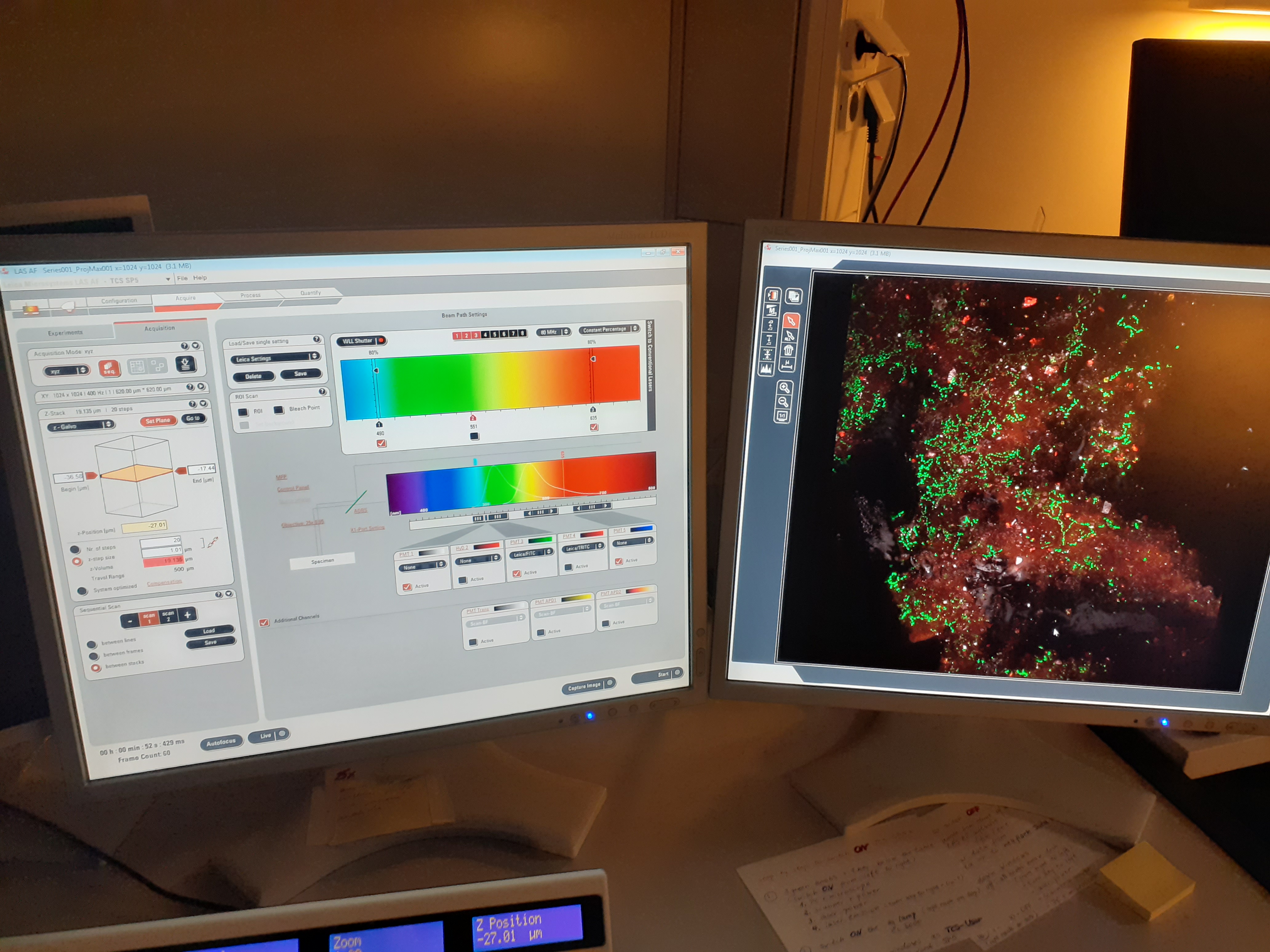
Confocal Laser Scanning Microscopy (CLSM)
Confocal Laser Scanning Microscopy (CLSM) is a well-established technique for visualization of microbial aggregates and biofilms. The major point is that it is a nondestructive approach for studying biofilms allowing three-dimensional sectioning of fully hydrated, living, thick microbial communities. CLSM reduces the need of biofilm pretreatment such as disruption and fixation procedures which usually modify its structure and activity.

In UFZ Magdeburg we have a Leica TCS SP5 X. CLSM allows multichannel imaging of cellular and extracellular constituents and in combination with fluorescent in situ hybridization techniques offers quantitative information on a wide variety of variables including cell numbers, cell area, and biofilm thickness. With the use of specific fluorochromes up to five different structural variables can be recorded simultaneously or sequentially. The two-dimensional digital image series recorded can be examined and analyzed.
If you are interested in visualising the structural components of biofilms with CLSM, do not hesitate to contact us.
Contact
Examples of CLSM applications:
- Michler-Kozma, D. N., Neu, T. R., & Gabel, F. (2021). Environmental conditions affect the food quality of plastic associated biofilms for the benthic grazer Physa fontinalis. Science of The Total Environment, 151663.
- Lawrence, J. R., Swerhone, G. D., & Neu, T. R. (2019). Visualization of the sorption of nickel within exopolymer microdomains of bacterial microcolonies using confocal and scanning electron microscopy. Microbes and environments, ME18134.
- Vosshage, A. T., Neu, T. R., & Gabel, F. (2018). Plastic alters biofilm quality as food resource of the freshwater gastropod Radix balthica. Environmental science & technology, 52(19), 11387-11393.
- Möhle, R. B., Langemann, T., Haesner, M., Augustin, W., Scholl, S., Neu, T. R., ... & Horn, H. (2007). Structure and shear strength of microbial biofilms as determined with confocal laser scanning microscopy and fluid dynamic gauging using a novel rotating disc biofilm reactor. Biotechnology and bioengineering, 98(4), 747-755.
- Lawrence, J. R., & Neu, T. R. (1999). [9] Confocal laser scanning microscopy for analysis of microbial biofilms. Methods in enzymology, 310, 131-144.
- Neu, T. R., & Lawrence, J. R. (1997). Development and structure of microbial biofilms in river water studied by confocal laser scanning microscopy. FEMS Microbiology Ecology, 24(1), 11-25.
